

Cerebral Amyloid Angiopathy after SARS-CoV-2 Infection
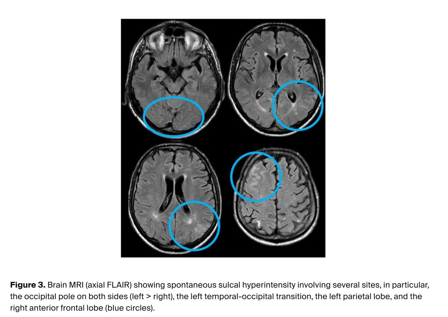
Spike protein driven hemorrhages in leptomeninges visible on MRI

By Peter A. McCullough, MD, MPH
A patient recently consulted me concerning the diagnosis of cerebral amyloid angiopathy given to him after an MRI was done at a prestigious university medical center. As expected he is genetically negative for inherited forms of amyloidosis. The patient was unvaccinated but did have severe, prolonged COVID-19 illness early in the pandemic. He is 67 years old and asymptomatic, which is unusual since ~70% of CAA patients have neurocognitive decline.
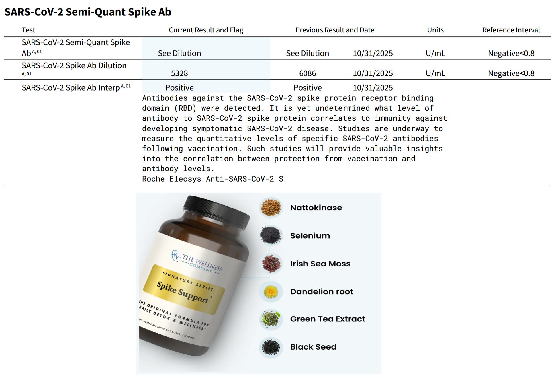
His anti-Spike antibodies were 6086 U/ml in October, 2025, declining to 5328 U/ml in February, 2026 on Spike Support from The Wellness Company. This has provided dissolution of the Spike protein without the additional anticoagulant effect of bromelain. Alter AI assisted in this medical review.
🧠 Cerebral Amyloid Angiopathy Following SARS-CoV-2 Infection
Cerebral amyloid angiopathy (CAA) represents a pathological state characterized by the deposition of β-amyloid (Aβ) peptides within the walls of small and medium-sized cerebral blood vessels, leading to vessel fragility, microhemorrhages, and inflammation. Traditionally viewed as a co-pathology of Alzheimer’s disease, CAA is increasingly recognized as an independent vascular neurodegenerative process. Recent research has revealed that SARS-CoV-2 infection—and particularly its Spike glycoprotein—can accelerate or precipitate amyloid deposition through inflammatory, endothelial, and molecular mimicry mechanisms. These findings suggest that the viral spike protein plays a key pathogenic role in the post-COVID cerebrovascular changes linked to amyloid pathology.
